

Abstract
Frequent infections are among the most common reasons for presentation to primary care and pediatric outpatient clinics. This review addresses the clinical evaluation and diagnostic approach to a child presenting with recurrent infections but otherwise clinically well and aims to help the clinician distinguish this child from one who requires further investigation. PubMed/MEDLINE, Scopus, and Web of Science were searched through 31 May 2026, with an update on 1 September 2026, to identify English- and Turkish-language literature on recurrent infections in children; sources were purposively selected by two authors, with priority given to guidelines, consensus documents, and cohort studies. Most recurrent infections in children occur in the context of physiological immune maturation and common exposure-related or host factors, including daycare or school attendance; large family size and household crowding; seasonal viral circulation; passive exposure to tobacco smoke; and atopic predisposition. In otherwise healthy children, infections are typically confined to the upper respiratory tract, are viral in nature, and are uncomplicated. Full recovery between episodes, normal growth and development, and a declining frequency of infections with age are expected. Four groups should be considered in the differential diagnosis: the healthy child with increased exposure, the child with atopic disease, the child with another chronic condition, and the child with an immune defect, whether inborn or secondary. Severe, unusual, or progressively worsening infections are warning signs, although published warning-sign lists have limited sensitivity and specificity. The primary goal of clinical evaluation is to distinguish the otherwise healthy child from the child requiring further investigation for inborn errors of immunity, secondary immunodeficiency, chronic disease, anatomical anomalies, cystic fibrosis, primary ciliary dyskinesia, or uncontrolled allergic disease. In the otherwise healthy child, family education, reduction of modifiable environmental risk factors, avoidance of unnecessary antibiotics and investigations, and structured follow-up are the priorities.
Keywords: Respiratory tract infections, recurrence, primary immunodeficiency diseases, differential diagnosis, child, primary health care
Introduction
Frequent infections in childhood commonly prompt presentation to family medicine and pediatric outpatient clinics. Parents often seek medical advice with concerns such as “Is my child’s immune system weak?” or “Is there an underlying serious disease?” However, a considerable proportion of children with frequent infections are immunologically normal.
Traditionally, children presenting with recurrent infections are grouped into four broad categories: the otherwise healthy child with increased exposure, the child with atopic disease, the child with another underlying chronic condition, and the child with an immune defect. Approximately half of these children are healthy and nearly one-third have an atopic constitution, while chronic disease and immunodeficiency together account for the remainder; in a Turkish series of 507 children presenting with recurrent infections to a pediatric infectious diseases and clinical immunology clinic, 52.3% were healthy, 29.2% had atopic disease, 7.9% had a chronic disorder and 10.7% had a primary immunodeficiency (). These proportions are largely derived from selected populations that were referred to secondary or tertiary care. In unselected primary care populations, the proportion of children with an underlying immune defect is considerably lower.
When evaluating a frequently ill child, it is more appropriate to focus not merely on the number of infections but on their characteristics, severity, anatomical localization, presence of complications, response to treatment, inter-episode clinical well-being, and the normality of the child’s growth and development. This is particularly important because frequent viral upper respiratory tract infections in preschool children may be observed more commonly in the context of daycare attendance, large families and household crowding (,).
Because recurrent respiratory infections (RRIs) may reflect either normal childhood exposure or an underlying disorder, the clinician must reassure and support the large majority of families whose child is well, while reliably recognizing the minority in whom further investigation is warranted (). This review is structured around that distinction.
Several reviews have addressed the child with recurrent infections (,,,). This review adds a discussion of post-pandemic considerations, a critical appraisal of warning-sign lists, and the integration of the 2025 ERS/ATS guideline on primary ciliary dyskinesia, the 2025 Joint Task Force practice parameter, and the 2024 IUIS classification, all set in the context of primary care in Türkiye. Its main sections follow the four categories outlined above.
Literature Search
This article is a narrative review. A narrative design was chosen because the question it addresses, namely, how to evaluate a child presenting with recurrent infections, is broad and clinically oriented. It spans several disciplines (general pediatrics, immunology, pulmonology, otorhinolaryngology, and allergy) and draws on heterogeneous evidence, including cohort studies, consensus statements, and clinical guidelines, which do not lend themselves to quantitative synthesis. The manuscript was self-assessed against the criteria of the Scale for the Assessment of Narrative Review Articles () ().
PubMed/MEDLINE, Scopus, and Web of Science were searched from inception to 31 May 2026 without restriction on publication year; the search was updated on 1 September 2026. Terms covering the scope of the review (“recurrent respiratory infections”, “recurrent infections”, “frequently ill child”, “inborn errors of immunity”, “primary immunodeficiency”, “secondary immunodeficiency”, “immunoglobulin deficiency”, “warning signs”, “cystic fibrosis”, “primary ciliary dyskinesia”, “bronchiectasis”, “allergic rhinitis”, “asthma”, “adenoid hypertrophy”, “immune debt” and “bacterial lysates”) were combined with “children”. Articles published in English or Turkish were eligible. Sources were purposively selected based on their clinical relevance to the review question. Priority was given to clinical guidelines, consensus documents, and prospective cohort studies, followed by systematic and narrative reviews. Studies confined to adult populations, conference abstracts, and case reports were not considered, nor were articles whose full text could not be obtained. The reference lists of selected articles were hand-searched for additional sources. Two authors identified and selected the literature independently, and disagreements were resolved by consensus. Additional sources addressing points raised during peer review were added during revision. Consistent with the narrative design, the search was not intended to be exhaustive, and the number of records at each stage was not reported.
Terminology and Definitions
The clinical entity described in Turkish as “sık hastalanan çocuk” overlaps with but is not identical to the entities the international literature calls RRIs and recurrent upper respiratory tract infections (R-URTIs) when episodes are restricted to the upper airway. RRI is a predominantly respiratory condition, usually operationalized with numerical criteria. A child who is frequently ill represents a broader presenting problem. It includes recurrent non-respiratory infections and children whose episode counts fall below any formal threshold but whose families still seek help. This review takes the broader presentation as its starting point. Because the evidence base is predominantly respiratory, the review draws mainly on the RRI literature and addresses recurrent infections at other sites in a separate section. There is no universally agreed numerical definition. A widely used criterion is eight or more documented airway infections per year in children under three years of age, or six or more per year in children older than three years of age, in the absence of an underlying pathological condition. An Italian inter-society consensus subsequently proposed an age-stratified definition, and a prospective primary-care cohort more recently proposed a pragmatic criterion for R-URTI: at least four upper respiratory episodes, each lasting four days or longer, within a six-month period, with intervals of complete well-being (,).
Recurrence of specific conditions is more clearly defined: acute otitis media is regarded as recurrent when there are three episodes within six months or four within twelve months, and infectious rhinitis when there are more than five episodes per year (). Recurrent pneumonia is generally defined as at least two episodes in one year, or three episodes ever, with radiographic clearing between episodes (). These thresholds are clinically more useful than a global infection count because they focus attention on the involved anatomical site.
How Should Normal Infection Frequency Be Evaluated?
There is no definitive threshold for the number of infections that should be considered “normal” in children. Age, daycare or school exposure, passive tobacco smoke exposure, seasonality, socioeconomic conditions, vaccination status, and the presence of atopic disease may all substantially influence the frequency of infections ().
In a German birth cohort followed to 12 years of age, parents reported a mean of 3.4 respiratory infections per year in the first two years of life, 2.3 in the preschool years and 1.1 at school age, with wide interindividual variation; the authors proposed upper limits of normal (mean plus two standard deviations) of 11, 8 and 4 episodes per year, respectively (). Daycare attendance and passive tobacco smoke exposure increase this burden, and large families and household crowding add to it in children who do not attend daycare (). Respiratory infection burden is highest in early childhood and generally decreases with age (,). Prospective cohort studies also show substantial interindividual variability in respiratory illness burden (,).
In otherwise healthy children, infections are usually mild to moderate, present as viral upper respiratory tract infections, resolve within the expected time frame, and children appear completely well between episodes. Symptoms such as fever, rhinorrhea, cough, and sore throat may recur; however, persistent fatigue, weight loss, growth failure, chronic diarrhea, recurrent pneumonia, or invasive bacterial infections are not expected between these episodes. Therefore, the number of infections alone is not an indicator of immunodeficiency; clinical findings and infection patterns are more decisive (,).
It has been estimated that approximately 25% of children under one year of age and at least 6% of children during the first six years of life experience recurrent respiratory tract infections, as a result of increased exposure to infectious agents during the early years of life, when immune functions are still maturing (,). In most of these children, the course is benign, and the frequency of episodes declines substantially as the children grow older, with the majority no longer affected by adolescence.
Category 1–The Frequently Ill but Otherwise Healthy Child
Who Is the Frequently Ill but Otherwise Healthy Child?
The frequently ill but otherwise healthy child is defined as one who has normal growth and development; age-appropriate weight and height percentiles; complete clinical well-being between infections; infections that are generally limited to the upper respiratory tract; recovery with appropriate treatment; and no history of severe or opportunistic infections. In these children, the onset of infections is often temporally associated with starting daycare or school or with increased seasonal viral circulation ().
Although the clinical picture may be distressing for families, in most cases it reflects frequent exposure to infectious agents rather than a pathological failure of the immune system. During the preschool period in particular, the immune system encounters numerous viral antigens to which it has not previously been exposed. This process is part of the normal immunological learning phase during childhood. If infections decrease with age, do not become more severe, and follow an uncomplicated course, the prognosis is generally favorable (). Reassuring features and warning signs are summarized in .
Table 1. Reassuring features and warning signs in the child presenting with recurrent infections
Domain | Reassuring features | Warning signs warranting further evaluation |
|---|---|---|
Growth and development | Weight and height on appropriate percentiles; normal development | Faltering growth, weight loss or developmental delay |
Condition between episodes | Completely well, active and feeding normally | Persistent fatigue, chronic diarrhea or unexplained recurrent fever |
Site of infection | Confined to the upper respiratory tract | Deep-seated or invasive infection (sepsis, meningitis, osteomyelitis, deep abscess) |
Anatomical pattern | Sites may vary within the upper respiratory tract | Repeated infection always at the same single site (suggests a local structural cause); severe infection at multiple sites (suggests a systemic immune defect) |
Causative organisms | Common respiratory viruses | Opportunistic or unusual organisms, treatment-resistant candidiasis, local or disseminated BCG infection or other complications following live vaccines |
Severity and course | Mild to moderate; resolves within the expected time | Severe, prolonged or progressively worsening episodes |
Response to treatment | Resolves with supportive care, or with oral antibiotics where indicated | Requires intravenous antibiotics to clear; fails to respond to appropriate therapy |
Trend with age | Frequency declines as the child grows older | Frequency or severity increases with age |
Timing of onset | Coincides with starting daycare or school | Onset in early infancy |
Family history | Unremarkable | Consanguinity, early infant death, known inborn error of immunity, unexplained autoimmunity |
Physical examination | Normal; tonsillar and lymphoid tissue present; mild to moderate eczema responding to standard treatment | Absent tonsillar or lymphoid tissue in the absence of previous surgery, hepatosplenomegaly, persistent lymphadenopathy, digital clubbing, severe, treatment-refractory eczema, particularly with recurrent skin infections, petechiae or markedly elevated IgE, oral candidiasis, non-healing wounds; delayed separation of the umbilical cord |
Laboratory findings, where obtained | Within age-specific reference ranges | Persistent lymphopenia or neutropenia; immunoglobulins low for age |
Compiled from references , , and . Warning signs indicate a need for further evaluation; they are not diagnostic criteria and should be applied alongside clinical judgment. BCG: Bacillus Calmette-Guérin
Risk Factors
The most important risk factors in frequently ill but otherwise healthy children are daycare or school exposure and atopy (). Crowded indoor environments facilitate the transmission of viral pathogens. Large families and household crowding similarly increase the infectious disease burden. Household tobacco smoke exposure is a preventable environmental factor that increases the risk of both upper and lower respiratory tract infections. Crowded living conditions and other environmental exposures may further increase opportunities for respiratory pathogen transmission ().
Post-pandemic Considerations
Since 2022, clinicians have encountered an atypical and often off-season resurgence of pediatric respiratory infections following the relaxation of non-pharmaceutical interventions introduced during the Coronavirus Disease-2019 pandemic. This rebound has been attributed to the concept of “immune debt” or “immunity gap”, that is, an enlarged pool of immunologically naive young children who missed early-life encounters with common pathogens (). The concept remains a matter of debate. Critics have cautioned that the term may be misread as implying lasting damage to children’s immune function, for which there is no evidence (), and recent reviews attribute the resurgence to a population-level immunity gap to specific pathogens rather than to altered childhood immune function (). What the concept describes, therefore, operates at the population level, not in the individual child. Nevertheless, it is clinically useful: it may explain an apparent increase in episode frequency without implying underlying pathology, and it provides the clinician with a reassuring point to discuss with anxious parents.
Category 2–Atopic Disease and Other Conditions That Mimic Recurrent Infection
Atopic diseases may increase the perception of frequent infections. Allergic rhinitis may be interpreted by families as a “constant infection” because of chronic nasal obstruction, postnasal drip, nocturnal cough, and recurrent sinusitis-like symptoms. However, in these children, the underlying problem is not always infection; rather, chronic mucosal sensitivity and increased secretions due to allergic inflammation may be responsible ().
Similarly, asthma, adenoid hypertrophy, gastroesophageal reflux, recurrent aspiration, chronic rhinosinusitis, and exposure to indoor or outdoor air pollution may all produce recurrent cough, rhinorrhea, or wheezing, which are repeatedly labeled and treated as infections. The pattern of antibiotic use is a useful indicator. Repeated courses without a clear clinical response may point to a non-infectious mimic instead of a bacterial etiology (,). Recognizing and treating the underlying atopic or anatomical condition frequently resolves what the family experiences as “constant infection” without further immunological investigation.
Category 3–The Child with an Underlying Chronic Condition
A distinct group of children with recurrent infections has neither normal immunity with high exposure nor an inborn error of immunity, but rather has an underlying non-immunological condition. This group is easily overlooked, yet it accounts for a clinically important proportion of referrals and often has a specific, treatable cause.
The location of the episodes is the most useful discriminator. Infection confined to one anatomical site points to a local structural or functional problem; severe infection at several sites points to a systemic immune defect (). In the first case the differential includes adenoid or tonsillar hypertrophy, chronic otitis media with effusion, anatomical obstruction of the sinuses, congenital lung malformation, retained foreign body, tracheoesophageal fistula, laryngeal cleft and bronchiectasis (,). In a recent Turkish series of 250 children with recurrent pneumonia, an underlying cause was identified in 89.6%; recurrence in the same area was associated mainly with asthma, right middle lobe syndrome and congenital lung or airway anomalies, and recurrence in different areas mainly with neuromuscular disease ().
Diffuse or lower respiratory tract involvement widens the differential diagnosis to include cystic fibrosis, primary ciliary dyskinesia, chronic aspiration secondary to swallowing dysfunction or neurological impairment, congenital heart disease with increased pulmonary blood flow, and tuberculosis in appropriate settings. Certain features help distinguish between them. Neonatal respiratory distress in a term infant, situs inversus, persistent rhinorrhea from the first weeks of life, and chronic wet cough all suggest primary ciliary dyskinesia. Failure to thrive with steatorrhea, nasal polyps or a history of meconium ileus points instead to cystic fibrosis (). None of these conditions are managed as normal variation or as an inborn error of immunity, which is why they need to be actively excluded.
The 2025 joint guideline of the European Respiratory Society and the American Thoracic Society bases the diagnosis of primary ciliary dyskinesia on a combination of tests. Measurement of nasal nitric oxide during velum closure is recommended alongside genetic testing and/or transmission electron microscopy. In younger children who cannot achieve velum closure, measurement during tidal breathing is an option, although it is less accurate. A normal nasal nitric oxide result does not exclude the diagnosis; therefore, the test cannot be used alone, and high-speed video microscopy and immunofluorescence may be added to the algorithm. No single test has complete sensitivity and specificity. A child with a strong clinical phenotype warrants referral to a specialist center even when one test result is normal ().
Recurrent Infections Outside the Respiratory Tract
The principle that applies to the respiratory tract applies elsewhere: infection repeatedly confined to one site suggests a local cause, whereas severe or unusual infection at several sites suggests an immune defect (). Recurrent urinary tract infection most often reflects a structural or functional abnormality of the urinary tract, such as vesicoureteral reflux, dysfunctional voiding or constipation; current guidance recommends specialist referral, with the choice and timing of renal imaging determined by the child’s age and the pattern of infection (). Immunological evaluation is warranted when urinary infections are accompanied by severe or unusual infections at other sites (). Recurrent deep skin or internal organ abscesses, particularly with Staphylococcus aureus, suggest a phagocyte defect such as chronic granulomatous disease, and recurrent skin infections accompanied by severe eczema suggest autosomal dominant hyper-IgE syndrome (,). Persistent oral candidiasis or fungal skin infections suggest a T-cell defect or chronic mucocutaneous candidiasis (,). Recurrent fever without an identifiable focus, occurring at regular intervals with complete well-being between episodes, points to an autoinflammatory disorder rather than recurrent infection. In children, periodic fever, aphthous stomatitis, pharyngitis and cervical adenitis syndrome and the hereditary recurrent fevers should be considered, and validated classification criteria are available for both (). Familial Mediterranean fever is frequent in Türkiye and should be considered early in this setting ().
Category 4–The Child with an Immune Defect
Secondary Immunodeficiency
Secondary (acquired) immunodeficiency is considerably more common than inborn errors of immunity and should be considered early in the evaluation, before or alongside extensive genetic investigation. Nutritional causes include protein-energy malnutrition and clinically significant micronutrient deficiencies; low vitamin D status has been associated with susceptibility to respiratory infection, although its causal contribution to secondary immunodeficiency remains unestablished. Protein is lost in nephrotic syndrome and in protein-losing enteropathy. Immunosuppressive therapy is an additional cause, most commonly involving systemic or repeated courses of corticosteroids or treatments for malignancy or autoimmune disease. Asplenia and functional hyposplenism, poorly controlled diabetes mellitus, chronic kidney or liver disease and human immunodeficiency virus infection complete the list (,,,).
A careful medication and nutritional history, together with an assessment of growth, should be obtained in every such evaluation and frequently identifies a reversible contributor. The first step is to correct or treat the cause. Whether further immunological evaluation is necessary depends on the degree of clinical suspicion that remains after the initial concern has been addressed.
When Should Inborn Errors of Immunity Be Suspected?
Antibody deficiencies are among the most frequently encountered inborn errors of immunity in clinical practice; however, the current classification of the International Union of Immunological Societies encompasses a much broader spectrum of immunological and immune-dysregulatory disorders ().
The proportions cited in the Introduction derive from children seen in specialist clinics and should not be extrapolated to primary care. Consanguineous marriage remains common in Türkiye: in the 2018 Turkish Demographic and Health Survey, approximately one in four marriages was consanguineous, although the proportion has declined in more recent marriage cohorts (). In a two-center Turkish registry, the estimated prevalence of primary immunodeficiency was 30.5 per 100,000 (). These findings suggest that regional differences in consanguinity and in the burden of inborn errors of immunity should be considered when interpreting data derived from other populations; however, referral-based or two-center estimates cannot be directly extrapolated to the primary care population of Türkiye.
Although frequent infections in healthy children are usually benign, the findings summarized in should raise suspicion for primary immunodeficiency or, in current terminology, inborn errors of immunity (,). Hypocalcemia in infancy, congenital heart defects, absence of the thymic shadow, and delayed separation of the umbilical cord are particularly important clues for syndromic immunodeficiencies (). Because BCG is given in early infancy in Türkiye, BCG lymphadenitis or disseminated BCG infection may be the first manifestation of severe combined immunodeficiency or Mendelian susceptibility to mycobacterial disease; in a series of 197 children with BCG complications and an inborn error of immunity, chronic granulomatous disease, severe combined immunodeficiency and Mendelian susceptibility to mycobacterial disease were the most common diagnoses (,).
In addition, infections that increase or become more severe with age, rather than decreasing as expected, should be considered pathological.
The 2025 inborn errors of immunity practice parameter of the Joint Task Force on Practice Parameters, issued jointly by the American Academy of Allergy, Asthma & Immunology, the American College of Allergy, Asthma and Immunology, and the Clinical Immunology Society, published in 2026, broadens the clinical triggers for investigation beyond infection alone. It recommends that an inborn error of immunity be considered in patients with recurrent, severe, or rare infections and in those presenting with autoinflammation, autoimmunity, severe atopy, atypical malignancy, bone marrow failure, or any combination of these features. It further recommends that a detailed family history be obtained, both to support the diagnosis and to identify affected relatives who remain undiagnosed ().
Warning-sign Lists and Their Limitations
The ten warning signs proposed by the Jeffrey Modell Foundation are the most widely disseminated screening aid for primary immunodeficiency and remain useful for raising awareness among non-specialists (). They should be applied with an understanding of their limitations. In a pediatric validation study, only a minority of the ten signs discriminated between children with and without a primary immunodeficiency; a positive family history, the need for intravenous antibiotics to clear infection, and failure to thrive performed best, whereas several of the infection-count criteria performed poorly (). It has consequently been argued that the list is neither sufficiently sensitive nor sufficiently specific to serve as a standalone screening instrument ().
A substantial proportion of patients with inborn errors of immunity present initially with non-infectious manifestations, including autoimmunity, immune dysregulation, lymphoproliferation, unexplained inflammation and malignancy, and not with recurrent infection alone (). The molecular spectrum of these disorders also continues to expand rapidly. The 2024 phenotypic classification of the International Union of Immunological Societies includes 559 inborn errors of immunity (), while the accompanying genotypic classification comprises 508 genes and 17 phenocopies (). The two figures differ because the phenotypic classification counts disorders, whereas the genotypic classification counts genes; variants in a single gene can cause more than one disorder, for example, through different modes of inheritance or through loss- vs. gain-of-function mechanisms. Warning-sign lists are best treated as an adjunct to clinical judgment and cannot replace it.
Clinical Evaluation
History Taking
A detailed history is central to the evaluation of the frequently ill but otherwise healthy child. The age at onset of infections, frequency, duration, seasonality, localization, fever pattern, antibiotic use, history of hospitalization, microbiological agents, complications, and clinical well-being between infectious episodes should be carefully elicited ().
The timing of daycare attendance, school environment, number of siblings, household tobacco smoke exposure, household crowding, sleep pattern, and nutritional status should also be evaluated. Vaccination history should be reviewed. Family history should include early infant death, recurrent infections, autoimmune disease, malignancy, consanguinity, and known immunodeficiency ().
Another important point is whether the episodes described as infections are truly infectious, since the conditions described in Category 2 can produce recurrent cough, rhinorrhea, or wheezing. Therefore, the question “How many times has the child used antibiotics?” is important, but so is “Were antibiotics truly indicated, and did the child respond to them?” Where available, documentation of previous episodes—clinic records, microbiological results and radiographs—may provide a more reliable basis for assessment than parental recall alone.
Physical Examination
The initial priorities during the physical examination are the child’s general appearance, growth and developmental status, and signs of chronic disease. Height, weight, and percentile curves should be assessed. In otherwise healthy children, growth and development are generally normal. Growth failure, poor weight gain, chronic diarrhea, persistent fever, hepatosplenomegaly, lymphadenopathy, skin abscesses, severe or treatment-refractory eczema, or oral candidiasis require further evaluation (,).
During an upper respiratory tract examination, tonsillar tissue, adenoid facies, nasal mucosa, allergic salute sign, transverse nasal crease, chronic nasal obstruction, and postnasal drip should be assessed. The lower respiratory tract examination should evaluate persistent wheezing, rales, respiratory effort, and chest deformities. In children with recurrent otitis media or sinusitis, an anatomical defect or allergic predisposition should be considered (,). The absence of tonsillar and lymphoid tissue in a child with recurrent infections is an important negative finding that should prompt evaluation for agammaglobulinemia, while digital clubbing suggests chronic suppurative lung disease.
Laboratory Evaluation
Extensive immunological testing is often unnecessary in children who appear clinically healthy, have normal growth, and experience mild infections limited to the upper respiratory tract. The decision to investigate should rest on clinical grounds, not on parental concern. Where anxiety persists despite a reassuring assessment, structured explanation and scheduled review are preferable to laboratory testing in the absence of clinical indications. Initial laboratory assessment may include complete blood count with differential, peripheral blood smear, absolute lymphocyte and neutrophil counts, serum immunoglobulin (Ig) levels, and, when indicated, complement levels (). Absolute lymphocyte and neutrophil counts should be carefully reviewed; findings such as lymphopenia, neutropenia, or eosinophilia may provide important clues regarding immunodeficiency subtypes. A stepwise approach is outlined in .
Table 2. Stepwise diagnostic evaluation of the child presenting with recurrent infections
Investigation | What it may identify |
|---|---|
Step 1—Initial evaluation, feasible in primary care | |
Full blood count with differential; absolute neutrophil and lymphocyte counts; peripheral blood smear | Persistent lymphopenia (cellular or combined defect), neutropenia, eosinophilia; Howell–Jolly bodies suggesting asplenia |
Serum IgG, IgA and IgM, interpreted against age-specific reference ranges | Predominantly antibody deficiency; transient hypogammaglobulinemia of infancy; selective IgA deficiency |
Structured review of growth, nutrition and medication history | Secondary immunodeficiency: malnutrition, micronutrient deficiency, corticosteroid or immunosuppressive therapy, protein-losing states |
Step 2—Targeted tests, selected according to the clinical pattern | |
Sweat chloride test | Cystic fibrosis |
Total and allergen-specific IgE; skin prick testing | Atopic disease mimicking recurrent infection |
Chest radiography; sinus imaging where indicated | Recurrent pneumonia, bronchiectasis, chronic rhinosinusitis; absent thymic shadow in infancy |
Age-appropriate HIV testing | Secondary immunodeficiency due to HIV infection |
Step 3—Second-line immunological tests, with specialist input | |
Vaccine-specific antibody titers before and after immunization, using protein and polysaccharide antigens | Specific antibody deficiency. Responses to polysaccharide antigens cannot be reliably interpreted below two years of age |
Lymphocyte subsets by flow cytometry | T-, B- and NK-cell defects; combined immunodeficiency |
C3, C4, CH50 and AH50 | Complement deficiencies |
Step 4—Advanced tests, performed in specialist centers | |
Dihydrorhodamine or nitroblue tetrazolium assay | Chronic granulomatous disease and other phagocyte defects |
Nasal nitric oxide measurement during velum closure, with genetic testing and/or transmission electron microscopy; high-speed video microscopy or immunofluorescence where available | Primary ciliary dyskinesia. A combination of tests is required; a normal nasal nitric oxide result does not exclude the diagnosis |
IgG subclass quantification | Selected antibody deficiencies |
Targeted gene panel or whole-exome sequencing | Molecular confirmation and classification of an inborn error of immunity |
HIV testing should be selected according to age and exposure history. Compiled from references , , , , and . Investigations should be selected on the basis of the history and physical examination, not ordered as an undirected screening panel; in the child who is well between episodes and growing normally, Step 1 is often sufficient. IgG subclass quantification is technically simple and widely available; it is placed in Step 4 not because of technical complexity but because an isolated low subclass level gains clinical significance mainly in the presence of recurrent infections, and its interpretation requires repeat measurement and assessment of specific antibody responses (,), which usually involves specialist input. AH50: Alternative pathway hemolytic complement activity, C3: Complement component 3, C4: Complement component 4, CH50: Total hemolytic complement activity, HIV: Human immunodeficiency virus, IgA: Immunoglobulin A, IgE: Immunoglobulin E, IgG: Immunoglobulin G, IgM: Immunoglobulin M, NK: Natural killer
Serum Ig concentrations must be interpreted against age-specific reference ranges. IgG reaches its physiological nadir at approximately three to six months of age, while IgA rises only slowly through childhood; interpretation against adult ranges is one of the most common sources of misdiagnosis. Several patterns recur in children who are otherwise well. Transient hypogammaglobulinemia of infancy is characterized by IgG levels below the age-specific reference range in an infant or toddler who generally has preserved specific antibody responses and normal growth. It is a retrospective diagnosis that can be confirmed only once Ig concentrations normalize, a process that may take several years; a proportion of children initially labeled in this way are subsequently found to have a persistent antibody deficiency. Serial measurement and clinical follow-up are essential, and management should be individualized according to the severity and burden of infections. Selective IgA deficiency is among the most common inborn errors of immunity and is often asymptomatic. It is diagnosed in patients older than four years with serum IgA below the laboratory limit of detection and normal serum IgG and IgM levels (). This age threshold is important because IgA rises slowly through early childhood, and low IgA values before this age are frequently transient. Clinicians should nevertheless be aware of its association with celiac disease and autoimmunity. Specific antibody deficiency, in which total Igs are normal but responses to polysaccharide antigens are impaired, is diagnosed by measuring vaccine-specific antibody titers before and after immunization. Responses to polysaccharide vaccines cannot be reliably interpreted below two years of age (,,).
Based on the working diagnosis, vaccine antibody responses, lymphocyte subset analysis, neutrophil function tests, sweat testing, chest radiography, sinus imaging, and advanced molecular or genetic investigations may be planned (,). These tests, however, should not be ordered as part of a broad, uncontrolled screening approach; rather, they should be selected based on clinical findings obtained through detailed history-taking and physical examination.
Molecular testing is not required for every otherwise healthy child with frequent infections; the indication for testing should be determined on the basis of clinical findings and the infection pattern. Where clinical suspicion is genuine, however, the expanding molecular spectrum outlined above underscores the importance of timely referral and appropriate genetic evaluation.
When to Refer
Referral to a pediatric immunologist is warranted when there is a family history of inborn errors of immunity or unexplained early infant death; when infections are severe, opportunistic, deep-seated, or require intravenous antibiotics; when there is failure to thrive, persistent lymphopenia or neutropenia, or unexplained autoimmunity; and when initial laboratory evaluation is abnormal after correction for age. Referral to a pediatric pulmonologist or otorhinolaryngologist is appropriate for chronic wet cough, recurrent infection confined to a single anatomical site, or suspicion of cystic fibrosis, primary ciliary dyskinesia, or an anatomical anomaly. In the child who is well between episodes, growing normally and has a normal initial evaluation, scheduled clinical review is preferable to either immediate referral or open-ended investigation (,,). The diagnostic approach is summarized in .
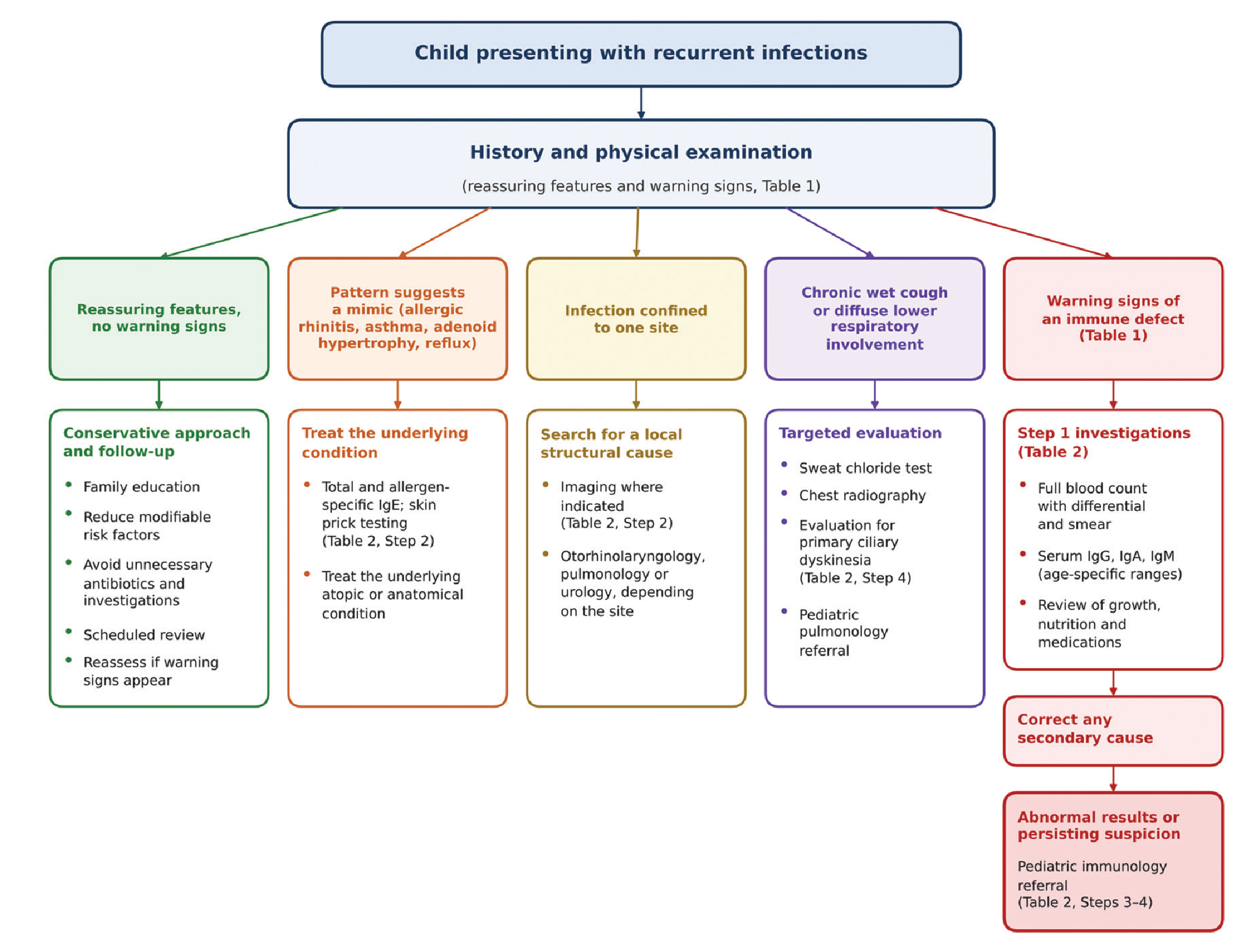
Diagnostic approach to the child presenting with recurrent infections
IgE: Immunoglobulin E
Management
Family Counseling
Families of frequently ill but otherwise healthy children should be informed that frequent viral infections during childhood, particularly during daycare and school years, are to be expected and that their frequency generally declines with age as immune maturation progresses and pathogen-specific immunity accumulates. Counseling should also emphasize avoiding unnecessary antibiotics and investigations. This explanation should be delivered with an understanding of parental anxiety and should involve the family in the clinical follow-up plan ().
Antibiotics should be used only when clinical findings support a bacterial infection. In recurrent viral upper respiratory tract infections, supportive care, adequate fluid intake, fever control, nasal hygiene, sleep regulation, and reduction of environmental risk factors are the main priorities. Eliminating passive tobacco smoke exposure, practicing hand hygiene, ventilating indoor environments, reducing contact with sick individuals, and receiving age-appropriate vaccination may reduce the infectious burden. In children with findings suggestive of allergic rhinitis or asthma, controlling the underlying atopic disease may substantially reduce the perception of “frequent infection.”
Families should be clearly informed about when to seek medical attention. Prolonged fever, respiratory distress, poor general condition, inability to feed, dehydration, and any of the warning signs listed in require prompt assessment and further evaluation.
Preventive Measures and Immunomodulators: What Is the Evidence?
Families frequently ask whether an “immune-boosting” preparation should be given, and a clear answer helps prevent both unnecessary expenditure and unrealistic expectations. The 2021 inter-society consensus, which graded the available evidence using the Grading of Recommendations Assessment, Development and Evaluation methodology, concluded that the evidence did not support the routine use of bacterial lysates such as OM-85, synthetic immunomodulators such as pidotimod, probiotics, trace elements, or vitamin C for the prevention of RRIs. Bacterial lysates and pidotimod may still be considered for selected children with a high frequency of episodes, considering the cost–benefit ratio. Routine vitamin D supplementation is not recommended for this indication, but supplementation is appropriate in individuals with deficiency or risk factors for deficiency. Age-appropriate immunization, including annual influenza vaccination, remains recommended, together with elimination of tobacco smoke exposure ().
Study Limitations
This article is a narrative review rather than a systematic review and does not present new, original, or epidemiological data. Although the cited literature was selected independently by two authors, narrative synthesis does not follow a prespecified protocol and remains subject to selection bias. Sources were selected purposively rather than through systematic screening with formal quality appraisal, and articles whose full text could not be obtained were not considered; relevant evidence may therefore have been missed. No single numerical threshold for “normal” infection frequency can be provided, since this varies with patient-specific and environmental factors. The review outlines a general clinical framework; it does not provide a universally applicable algorithm for advanced genetic and molecular testing. The proportions cited for the diagnostic categories derive largely from referral populations, which limits their generalizability to primary care.
Conclusion
This review combines recent guidelines and classifications with a critical view of warning-sign lists to offer a practical framework for primary care. The frequently ill but otherwise healthy child represents a common clinical scenario in pediatric and primary care practice. The key determinants in the evaluation are the severity, localization, complications, response to treatment, clinical well-being between episodes, and growth and developmental status. In children with viral infections limited to the upper respiratory tract and with normal growth and development, the priorities are family education, reduction of environmental risk factors, prevention of unnecessary antibiotic use, and regular clinical follow-up. Severe, unusual, or progressively worsening infections or infections involving multiple anatomical sites require further evaluation for inborn errors of immunity, while infections confined to a single anatomical site should prompt a search for a local structural cause; secondary causes of immunodeficiency should also be considered and addressed before or alongside advanced genetic investigation.
Ethics
Ethics Committee Approval: As this study is a review article based on previously published literature and does not involve human or animal subjects, ethics committee approval was not required.
Informed Consent: Not required.
Footnotes
Authorship Contributions
Concept: Ö.K., N.C.K., Design: Ö.K., N.C.K., Data Collection or Processing: Ö.K., N.C.K., Analysis or Interpretation: Ö.K., N.C.K., Literature Search: Ö.K., N.C.K., Writing: Ö.K., N.C.K.
Conflict of Interest
No conflict of interest was declared by the authors.
Financial Disclosure
The authors declared that this study received no financial support.
References
Sütçü M, Acar M, Aktürk H, Hançerli-Törün S, Salman N, Somer A. Recognizing immunodeficiency in children with recurrent infections: what are the predictive factors? Turk J Pediatr. 2016;58:609-615.
Cardinale F, Zuccarino F, Serio C, et al. Recurrent respiratory infections in children: new perspectives. Glob Pediatr. 2024;8:100105.
Duse M, Leonardi L, Zicari AM, De Castro G, Indinnimeo L. Risk factors for upper airway diseases. Int J Immunopathol Pharmacol. 2010;23:13-15.
Chiappini E, Santamaria F, Marseglia GL, et al. Prevention of recurrent respiratory infections. Ital J Pediatr. 2021;47:211.
Alkhater SA. Approach to the child with recurrent infections. J Family Community Med 2009;16:77-82.
Sütçü M, Kara M, Somer A. Approach to the frequently ill child. Çocuk Derg. 2018;18:47-51.
Reust CE. Evaluation of primary immunodeficiency disease in children. Am Fam Physician. 2013;87:773-778.
Reilly L, Emonts M. Recurrent or unusual infections in children: when to worry about inborn errors of immunity. Ther Adv Infect Dis. 2023;10:20499361231162978.
Baethge C, Goldbeck-Wood S, Mertens S. SANRA-a scale for the quality assessment of narrative review articles. Res Integr Peer Rev. 2019;4:5.
Corsello A, Milani GP, Picca M, et al. Recurrent upper respiratory tract infections in early childhood: a newly defined clinical condition. Ital J Pediatr. 2024;50:30.
Montella S, Corcione A, Santamaria F. Recurrent pneumonia in children: a reasoned diagnostic approach and a single centre experience. Int J Mol Sci. 2017;18:296.
Grüber C, Keil T, Kulig M, Roll S, Wahn U, Wahn V; MAS-90 Study Group. History of respiratory infections in the first 12 yr among children from a birth cohort. Pediatr Allergy Immunol. 2008;19:505-512.
Toivonen L, Karppinen S, Schuez-Havupalo L, et al. Burden of recurrent respiratory tract infections in children: a prospective cohort study. Pediatr Infect Dis J. 2016;35:e362-e369.
de Vries E, van Hout RWNM. Respiratory symptoms in post-infancy children. A dutch pediatric cohort study. Front Pediatr. 2020;8:583630.
Subbarayan A, Colarusso G, Hughes SM, et al. Clinical features that identify children with primary immunodeficiency diseases. Pediatrics. 2011;127:810-6.
Cohen R, Levy C, Rybak A, Angoulvant F, Ouldali N, Grimprel E. Immune debt: recrudescence of disease and confirmation of a contested concept. Infect Dis Now. 2023;53:104638.
American Association of Immunologists. AAI statement on the concept of “immunity debt” [Internet]. 2023 Aug 29. Available from: https://www.aai.org/AAISite/media/Public_Affairs/Letters-Comments/AAI-Statement-on-Immunity-Debt-August-2023_Final.pdf
Nygaard U, Holm M, Rabie H, Rytter M. The pattern of childhood infections during and after the COVID-19 pandemic. Lancet Child Adolesc Health. 2024;8:910-920.
de Benedictis FM, Bush A. Recurrent lower respiratory tract infections in children. BMJ. 2018;362:k2698.
Özçelik T, Can Oksay S, Girit S. Diagnostic approach to the etiology of recurrent pneumonia in children. Pediatr Pulmonol. 2024;59:3650-3659.
Shoemark A, Goutaki M, Kinghorn B, et al. European Respiratory Society and American Thoracic Society guidelines for the diagnosis of primary ciliary dyskinesia. Eur Respir J. 2025;66:2500745.
National Institute for Health and Care Excellence. Urinary tract infection in under 16s: diagnosis and management. NICE guideline NG224. London: NICE; 2022.
Gattorno M, Hofer M, Federici S, et al. Classification criteria for autoinflammatory recurrent fevers. Ann Rheum Dis. 2019;78:1025-1032.
Tunca M, Akar S, Onen F, et al. Familial Mediterranean fever (FMF) in Turkey: results of a nationwide multicenter study. Medicine (Baltimore). 2005;84:1-11.
Herman KE, Tuttle KL. Overview of secondary immunodeficiency. Allergy Asthma Proc. 2024;45:347-354.
Chinen J, Shearer WT. Secondary immunodeficiencies, including HIV infection. J Allergy Clin Immunol. 2010;125:S195-203.
Bousfiha AA, Jeddane L, Moundir A, et al. The 2024 update of IUIS phenotypic classification of human inborn errors of immunity. J Hum Immun. 2025;1:e20250002.
Koç İ. Changes in the prevalence of consanguineous marriages in Türkiye and identification of resistant groups: analyses by marriage cohorts from the 2018 Türkiye Demographic and Health Survey. Turk J Public Health. 2022;20:423-438.
Kilic SS, Ozel M, Hafizoglu D, Karaca NE, Aksu G, Kutukculer N. The prevalences [correction] and patient characteristics of primary immunodeficiency diseases in Turkey--two centers study. J Clin Immunol. 2013;33:74-83.
Fazlollahi MR, Goudarzi A, Nourizadeh M, et al. Complications of the Bacillus Calmette-Guerin vaccine as an early warning sign of inborn errors of immunity: a report of 197 patients. Front Immunol. 2024;15:1477499.
Orange JS, Chinen J, Horner CC, et al. 2025 Inborn errors of immunity practice parameter: Guidance from the Joint Task Force on Practice Parameters, the American Academy of Allergy, Asthma & Immunology (AAAAI), the American College of Allergy, Asthma and Immunology (ACAAI) and the Clinical Immunology Society (CIS). Ann Allergy Asthma Immunol. 2026;136:426-493.e1. Erratum in: Ann Allergy Asthma Immunol. 2026;S1081-1206(26)00390-X.
Jeffrey Modell Foundation. 10 warning signs of primary immunodeficiency in children [Internet]. Accessed August 31, 2026. Available from: https://info4pi.org
Arkwright PD, Gennery AR. Ten warning signs of primary immunodeficiency: a new paradigm is needed for the 21st century. Ann N Y Acad Sci. 2011;1238:7-14.
Thalhammer J, Kindle G, Nieters A, et al. Initial presenting manifestations in 16,486 patients with inborn errors of immunity include infections and noninfectious manifestations. J Allergy Clin Immunol. 2021;148:1332-1341.e5.
Poli MC, Aksentijevich I, Bousfiha AA, et al. Human inborn errors of immunity: 2024 update on the classification from the International Union of Immunological Societies Expert Committee. J Hum Immun. 2025;1:e20250003.
European Society for Immunodeficiencies. ESID Registry: working definitions for clinical diagnosis of PID [Internet]. Accessed 2026 Oct 18. Available from: https://esid.org/layout/set/print/content/download/15138/418138/file/ESIDRegistry_ClinicalCriteria.pdf
Koçaoğlu M, Koçaoğlu BE, Erol Aytekin S, et al. Clinical and laboratory evaluation of Turkish children with IgG subclass deficiency. Pediatr Neonatol. 2023;64:38-45.
