

To the Editor,
Osteopoikilosis is a rare, benign, and mostly asymptomatic sclerosing bone dysplasia. It is usually detected incidentally on plain radiographs obtained for other reasons and is typically characterized by symmetrical, well-defined, small, round-to-oval, juxta-articular sclerotic foci (, , ). When diffuse involvement is present, it may pose an important differential diagnostic challenge in clinical practice because it can mimic osteoblastic metastasis (). In addition, loss-of-function mutations in the LEMD3 gene have been reported to be associated with osteopoikilosis, Buschke–Ollendorff syndrome, and melorheostosis ().
A 33-year-old woman presented with left shoulder pain that had been intermittent for an extended period and had become more prominent over the preceding three months. She had been evaluated at different clinics for possible malignancy after bone lesions were observed on earlier radiographs. On physical examination, tenderness of the left biceps tendon and painful myofascial bands in the upper trapezius and levator scapulae muscles were noted. These clinical findings were considered sufficient to explain the patient’s shoulder symptoms, whereas the radiographic bone lesions were interpreted as incidental findings rather than the direct cause of pain. The neurological examination was normal, and no skin lesions were observed.
Plain radiographs revealed numerous symmetrical, juxta-articular, well-defined, homogeneous, round-to-oval, sclerotic foci around the bilateral humeral heads, wrists and carpal bones, metacarpals and phalanges, distal femurs, pelvis, bilateral hips, and ankles, with a distribution predominantly oriented toward adjacent joints (). The cortical contours were preserved, and no osseous destruction or periosteal reaction was observed. Bone scintigraphy, performed for the differential diagnosis of osteoblastic metastasis, was normal. The absence of increased tracer uptake, corresponding to a “cold” scintigraphic pattern, supported the diagnosis of osteopoikilosis and helped exclude osteoblastic metastases, which typically demonstrate increased uptake as “hot” lesions. Abdominopelvic ultrasonography showed no findings suggestive of malignancy. Erythrocyte sedimentation rate, C-reactive protein, complete blood count, renal and liver function tests, serum calcium, phosphorus, magnesium, bone-specific alkaline phosphatase, parathyroid hormone, and calcitonin levels were within normal limits; only the vitamin D level was low.
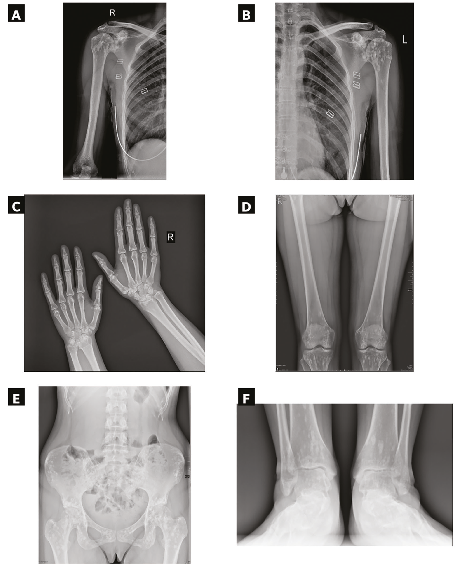
Based on the clinical, laboratory, and radiological findings, the patient was diagnosed with osteopoikilosis. The shoulder pain was considered more likely to be related to concomitant myofascial and tendinous pathology rather than directly to osteopoikilosis. A marked reduction in pain was observed at the first-month follow-up after medical treatment, physical therapy modalities, stretching exercises, dry needling, and vitamin D replacement. No additional clinical or imaging findings suggestive of malignancy were detected during follow-up.
Osteopoikilosis is generally a benign condition that does not require treatment; however, recognition of this entity is important because it may be confused with metastatic disease. The symmetrical, small, well-defined, enostosis-like, and periarticular distribution of the lesions, together with the absence of osseous destruction, periosteal reaction, and increased scintigraphic activity, is helpful in establishing the diagnosis (, , , , ). In contrast, osteoblastic metastases typically demonstrate increased radiotracer uptake on bone scintigraphy, making the technique particularly useful when diffuse sclerotic lesions raise concern for malignancy. Adherence to patient consent and transparent reporting principles is also important in case reports (). Osteopoikilosis should be considered in the differential diagnosis of diffuse sclerotic bone lesions. When pain is present, concomitant myofascial or tendinous causes should be evaluated separately.
Footnotes
Ethics
Informed Consent: Written informed consent was obtained from the patient for participation in this report and for the publication of clinical data and images.
Conflict of Interest: No conflict of interest was declared by the author.
Financial Disclosure: The author declared that this study received no financial support.
References
Woyciechowsky TG, Monticielo MR, Keiserman B, Monticielo OA. Osteopoikilosis: what does the rheumatologist must know about it? Clin Rheumatol. 2012;31:745-748.
Ozdemirel AE, Cakit BD, Erdem HR, Koc B. A rare benign disorder mimicking metastasis on radiographic examination: a case report of osteopoikilosis. Rheumatol Int. 2011;31:1113-1116.
Benli IT, Akalin S, Boysan E, Mumcu EF, Kiş M, Türkoğlu D. Epidemiological, clinical and radiological aspects of osteopoikilosis. J Bone Joint Surg Br. 1992;74:504-506. Erratum in: J Bone Joint Surg [Br]. 1994;76:683.
Hellemans J, Preobrazhenska O, Willaert A, et al. Loss-of-function mutations in LEMD3 result in osteopoikilosis, Buschke-Ollendorff syndrome and melorheostosis. Nat Genet. 2004;36:1213-1218.
Serdaroğlu M, Capkin E, Uçüncü F, Tosun M. Case report of a patient with osteopoikilosis. Rheumatol Int. 2007;27:683-686.
Paraskevas G, Raikos A, Stavrakas M, Spanidou S, Papaziogas B. Osteopoikilosis: a case report of a symptomatic patient. J Radiol Case Rep. 2009;3:38-43.
Gagnier JJ, Kienle G, Altman DG, et al. The CARE guidelines: consensus-based clinical case report guideline development. J Clin Epidemiol. 2014;67:46-51.
