

What is known on this subject?
A double-contour sign on chest radiography is characterized by two curvilinear densities on the cardiac site, often visualized as a retrocardiac opacity.
What this clinical images adds?
A double-contour sign on the cardiac site on chest radiography may indicate giant hiatal hernia.
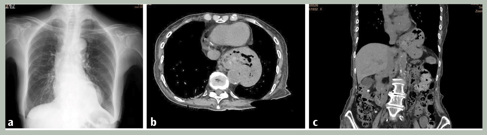
HH is classified into four types based on the position of the gastroesophageal (GE) junction, extent of the herniated stomach, and herniation of abdominal organs other than the stomach. In type I HH, also known as sliding hernia, the GE junction migrates into the mediastinum. In type II HH, also called paraesophageal hernia, the GE junction is in its normal position, but the gastric fundus herniates through the hiatus along its side. Type III HH is a combination of type I and II HH. In this type, the stomach protrudes through the hiatus, and the GE junction is displaced. Type IV HH is characterized by herniation of the stomach with other organs, such as colon, small intestine, spleen, and pancreas, via a large defect in the diaphragm (,). Although definitions vary, a giant HH is characterized by herniation of >30 to 50% of the stomach (). Therefore, the patient’s condition was categorized as type III giant HH.
In the current case, based on the left-sided double-contour sign on the cardiac site, a diagnosis of type III giant HH was made. The left-sided double-contour sign can be caused by pulmonary diseases (e.g., intralobular sequestration and endobronchial carcinoid tumor) and left ventricular masses (e.g., cardiac angiosarcoma and left ventricular pseudoaneurysm). Esophageal varices and mediastinal lipomatosis can lead to right- or left-sided double-contour signs. Extramedullary hematopoiesis can also result in a bilateral double-contour sign ().
Several cases of type III giant HH diagnosed based on the double-contour sign have been reported (,). However, these cases involve bilateral double-contour signs, not, left- or right-sided double-contour signs alone. In the current case, the right border of the type III giant HH overlapped the spine. Therefore, the right-sided double-contour sign could not be found on chest radiography.
In conclusion, clinicians should be aware that a double-contour sign on the cardiac site on chest radiography may indicate type III giant HH.
Footnotes
Ethics
Informed Consent: Informed consent was signed by the patient for this report.
Financial Disclosure: The author declared that this study received no financial support.
References
Banimostafavi ES, Tayebi M. Large hiatal hernia with pancreatic body herniation: case-report. Ann Med Surg (Lond). 2018;28:20-22.
Patel S, Shahzad G, Jawairia M, Subramani K, Viswanathan P, Mustacchia P. Hiatus hernia: a rare cause of acute pancreatitis. Case Rep Med. 2016;2016:2531925.
Mitiek MO, Andrade RS. Giant hiatal hernia. Ann Thorac Surg. 2010;89:S2168-S2173.
Gönlügür U, Yılmaz M, Reşorlu M, Kapıcıbaşı HO. Bilateral double contour on the cardiac borders and a review of the literature. Troia Med J. 2022;3:30-32.
Hirata K, Shimabukuro Y, Takahashi T, Wake M. Syncope due to intracavitary left ventricular obstruction secondary to giant esophageal hiatus hernia. Am J Med Case Rep. 2017;5:89-93.
